Scientists finally solved how H5N1 bird flu hid in dairy cows
How H5N1 Bird Flu Hid in US Dairy Cows: Unraveling the Molecular, Receptor, and Epidemiological Mechanisms Behind the Silent Outbreak
Executive Summary
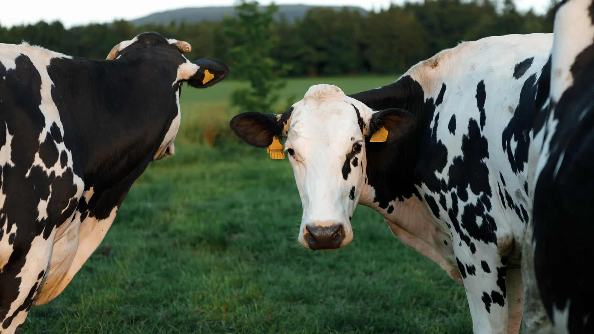 Scientists have finally solved the mystery of why H5N1 bird flu behaved so differently when it jumped into U.S. dairy cattle. Credit: Shutterstock
When H5N1 highly pathogenic avian influenza (HPAI) was first detected in US dairy cattle in early 2024, it confounded expectations. Highly pathogenic avian influenza viruses are, by definition, supposed to be devastating — killing chickens, turkeys, and wild birds in waves of conspicuous mortality. Instead, this virus slipped quietly into the udders of dairy cows, producing mastitis rather than mass death, spreading through routes distinct from conventional respiratory droplet transmission, and infecting hundreds of herds across at least 17 states before anyone fully understood what was happening.
A convergence of recent research has now solved this puzzle. The answer is not a single trick but a layered set of adaptations: a polymerase gene mutation (PB2 M631L) that unlocked efficient replication in bovine cells; a hemagglutinin mutation (HA T199I) combined with a uniquely favorable N-linked glycan architecture in mammary tissue that redirected the virus away from the respiratory tract and into the udder; and an atypical clinical presentation dominated by mastitis with high subclinical rates that allowed the virus to spread beneath the radar of routine veterinary surveillance. Together, these mechanisms explain how a virus expected to announce itself through mass mortality instead whispered through the American dairy industry.
Introduction: An Outbreak That Should Not Have Been Possible
For decades, the prevailing assumption among influenza virologists was that H5N1, while devastating to birds, would struggle to establish itself in mammals without sweeping reassortment or dramatic receptor-switching events. Cattle, in particular, were not considered a natural reservoir for influenza A viruses. Yet beginning in early 2024, the B3.13 genotype of H5N1 began appearing in dairy herds across the United States — eventually spreading to more than 1,000 herds across 17–18 states by mid-2026, with California alone accounting for roughly 75% of confirmed cases (Globe and Mail; USDA APHIS; CIDRAP).
What made this outbreak so unusual — and so dangerous — was its silence. Highly pathogenic avian influenza viruses typically kill 90–100% of infected chickens within days. In dairy cattle, the same virus produced a clinical picture dominated by thickened, discolored milk, decreased feed intake, and mild respiratory signs. Many infected cows showed no symptoms at all. A seroprevalence study found that 89.4% of cows in an affected herd (570 of 637) had antibodies to H5N1, yet 76.1% of those seropositive animals were subclinically infected — meaning they carried the virus without showing any visible signs of illness. Only around 15% of antibody-positive animals showed clinical disease, and the whole-herd clinical rate was 20% (777 of 3,876 adult cows) (Nature Communications). This high ratio of subclinical to clinical infection is precisely what allowed the virus to spread undetected across the country.
The question that has consumed researchers since the outbreak began is: how? How did an avian influenza virus learn to replicate in bovine cells, target the mammary gland instead of the lungs, and produce a clinical picture so atypical that it was mistaken for routine udder health problems? Recent studies have now provided a remarkably complete answer.
Part 1: The Polymerase Adaptation — Unlocking Replication in Bovine Cells
The Central Mutation: PB2 M631L
Two independent studies published in Nature Communications converge on the same central finding: a single mutation in the PB2 gene — PB2 M631L — was the critical adaptation that allowed avian-origin H5N1 to replicate efficiently in mammalian cells (Nature Communications 1; Nature Communications 2).
This mutation was found in essentially all cattle-derived H5N1 sequences, making it a defining feature of the bovine-adapted virus. Its functional impact was striking: the mutation enhanced polymerase activity in mammalian cells by 23.3-fold. The mechanistic explanation lies in the structural interface between the viral polymerase and a host protein called ANP32A. Most avian ANP32A proteins contain a 33-amino-acid insertion that the avian polymerase has evolved to interact with. Bovine ANP32A lacks this insertion — it is shorter. The PB2 M631L mutation enables the avian virus polymerase to productively interact with this shorter bovine ANP32A protein, unlocking efficient viral replication in bovine cells.
This is not merely a curiosity of bovine virology. The same mutation also enhances replication in human airway epithelial cultures, underscoring its zoonotic significance. The molecular bridge between bovine and human infection is, in effect, already partially built.
Supporting Polymerase Mutations
PB2 M631L did not act alone. Additional polymerase mutations contributed to full mammalian adaptation:
- PA K497R was found in the majority of cattle sequences and co-adapted with PB2 M631L to optimize polymerase function in mammalian cells.
- PB2 A129T, V495I, and V649I collectively raised polymerase activity to match cow-adapted efficiency.
- PB2 E627K and D740N are emerging substitutions that continue to increase replicative ability in mammals — a warning of further adaptation.
An important distinction: the B3.13 genotype itself arose from prior reassortment between Eurasian H5N1 and North American low pathogenic avian influenza (LPAI) viruses, which shaped its genome constellation before it ever entered cattle. However, the specific cattle-adaptive mutations — PB2 M631L and PA K497R — were not acquired through reassortment. They arose through selection within the cattle host after spillover, as the virus adapted to replicate efficiently in bovine cells. The virus did not inherit these adaptations from a pre-existing reservoir; it evolved them under pressure in its new mammalian host.
Historical Context
PB2 M631L is not a wholly new threat. It previously circulated in avian and human H5N1 strains during 2013–2014 outbreaks in Cambodia and Vietnam, and first emerged in Thailand as early as 2005. Its appearance in cattle represents the realization of a zoonotic risk that has been latent for nearly two decades. The fact that this mutation had been circulating in avian and human populations for years without causing a mammalian outbreak suggests that additional factors — including the receptor-binding adaptations described below — were necessary for the virus to establish itself in dairy cattle.
Part 2: The Receptor-Binding and Tissue-Tropism Adaptations — Why the Virus Targeted the Udder
The Key Mutation: HA T199I
A separate but equally critical adaptation occurred in the hemagglutinin (HA) protein. A single mutation — T199I — emerged in late 2023, just before the dairy cow outbreak began (Nature Communications).
T199I is located outside the canonical receptor-binding site but increases flexibility within the receptor-binding subdomain (RBS). This increased flexibility enables the virus to bind a broader range of glycans bearing terminal α2,3-linked sialic acids. The expanded binding breadth allowed the virus to effectively target the abundant α2,3 sialic acid-linked glycans in dairy cow mammary tissue.
This is a subtle but important distinction. The virus did not switch its receptor preference from avian-type (α2,3) to human-type (α2,6) — a change that would have raised immediate pandemic alarms. Instead, it broadened its ability to bind a wider variety of α2,3-linked glycans, which happen to be abundant in bovine mammary tissue.
Preserved Avian-Type Receptor Specificity
The bovine H5N1 virus retained strong specificity for avian-type (α2,3-linked) sialic acid receptors rather than adapting to human-type (α2,6-linked) receptors. Santos et al. demonstrated that the bovine H5N1 virus binds poorly to glycans terminating in α2,6 sialic acids — the receptors abundant in the human upper respiratory tract (Nature).
There is some disagreement in the literature on this point. One study from CMMR reported that bovine H5N1 HA can bind both α2,3 and α2,6 sialic acid receptors to some degree (CMMR). However, the more rigorous glycan-array analyses by Santos et al. indicate that the dominant specificity remains avian-type. This preserved avian-type receptor preference is key to understanding both how the virus hid in dairy cattle and why it has not yet efficiently adapted for human transmission — though any future mutation shifting toward stronger α2,6 binding would dramatically increase pandemic potential.
N-Linked Sialic Acid Receptors: The Critical Determinant of Udder Tropism
A breakthrough study led by Suresh Kuchipudi at the University of Pittsburgh School of Public Health, in collaboration with glycomics expert Lauren E. Pepi of Harvard Medical School and published in Science Advances, identified the precise molecular basis for the virus's mammary tropism (ScienceDaily).
Using binding experiments, glycan staining, and ultra-high-resolution imaging, the researchers mapped the detailed glycan architecture of bovine tissues. They discovered that H5N1 binds specifically to N-linked sialic acid receptors (as opposed to O-linked sialic acid receptors). These N-linked sialic acid receptors are concentrated throughout udder tissue but are nearly absent in bovine airway tissue. This stark tissue-distribution difference made mammary glands a “perfect breeding ground” for the virus, explaining why infected cows developed severe necrotizing mastitis rather than respiratory illness.
This finding adds a crucial new layer to the receptor story: it is not merely that avian-type α2,3 sialic acid receptors are present in mammary tissue, but that the specific N-linked presentation of those receptors — abundant in the udder and absent from the airways — determines tissue tropism. The Kuchipudi team's framework also provides a tool for preemptively screening other animal species and tissues for H5N1 susceptibility, potentially preventing future surprises in how the virus manifests across different hosts.
Mammary Gland Receptor Abundance
A complementary study published in Emerging Infectious Diseases (July 2024) provided histological confirmation of mammary tropism (CDC EID). Dairy cattle mammary glands are rich in SA α2,3-gal receptors — the avian influenza virus-specific receptor type. H5N1 virus co-localizes with these avian-specific receptors in mammary gland tissue. The epitheliotropism of H5N1 within the mammary gland, combined with the abundance of SA α2,3-gal receptors, provided the mechanism that allowed the virus to establish infection in dairy cattle after its introduction from wild birds.
This receptor-based explanation is critical for containment strategies: it explains why milk became the primary vector for viral shedding and why premovement testing of dairy cattle is necessary.
Part 3: The Clinical Mechanism — Why the Virus Went Undetected
Mastitis Instead of Mortality
The B3.13 genotype H5N1 virus produced an atypical clinical picture for a highly pathogenic avian influenza. Instead of the severe respiratory disease and high mortality that define HPAI in birds, infected dairy cows exhibited:
- Mammary gland tropism rather than severe respiratory disease
- Mastitis and milk loss as primary symptoms
- Modest respiratory distress rather than high mortality
- Subclinical infections in the majority of animals
This clinical presentation was so unlike what veterinarians expected from H5N1 that early cases were likely mistaken for routine udder health problems — environmental mastitis, bacterial infections, or nutritional issues. The virus was not on anyone's radar as a cause of mastitis in dairy cattle.
Physiological Changes Precede Visible Signs
Even in cows that did become symptomatic, physiological changes (such as decreased rumination) preceded visible clinical signs, creating a window during which infected animals appeared normal but were already shedding virus. This subclinical shedding period is a well-known feature of many infectious diseases, but it is particularly dangerous when combined with a virus being transmitted through non-respiratory routes.
Transmission Routes
Because the virus localized to mammary tissue and was shed in milk, contaminated milking equipment was the leading early hypothesis for cow-to-cow transmission — bypassing the respiratory close-contact patterns typical of influenza spread. However, this picture has since become more complicated. A May 2026 experimental study (Lee et al., Nature Communications) found that H5N1 does not readily transmit between dairy cows via contaminated milking equipment or close contact under controlled conditions, and a separate 2026 paper proposes cross-nursing — calf-to-cow oral-to-mammary contact — as an alternative route. The precise mechanism of within-herd spread remains an active area of investigation. What is not in question is that the mammary tropism facilitated silent spread across hundreds of herds in multiple US states through routes distinct from conventional respiratory influenza.
This is a critical departure from how influenza is normally understood to spread. Influenza viruses are typically thought of as respiratory pathogens transmitted through droplets and aerosols. The bovine H5N1 outbreak demonstrated that an influenza virus can exploit entirely different transmission dynamics when its tissue tropism directs it to a non-respiratory site. This has implications far beyond dairy cattle: it suggests that other livestock species with similar mammary gland architecture could be vulnerable to similar silent outbreaks.
Part 4: The Epidemiological Context
Two distinct viral genotypes have been identified in the US outbreak (Globe and Mail):
- B3.13 genotype — circulating in dairy cattle, the lineage responsible for the bovine outbreak.
- D1.1 genotype — found in wild birds and poultry, distinct from the dairy lineage.
The D1.1 genotype is associated with more severe human cases, including the first severe human case in the United States — a Louisiana resident hospitalised in December 2024 following exposure to backyard poultry. On the same day, California declared a state of emergency, though that declaration was driven primarily by the rapid surge in B3.13 dairy cattle outbreaks within California (over 300 new herd detections in the preceding 30 days), not by the Louisiana D1.1 case. The B3.13 genotype, while responsible for the vast majority of dairy herd infections, has generally produced milder illness in humans — though this should not be interpreted as a sign of safety. The virus is evolving, and the distinction between genotypes may not remain stable.
Containment efforts have been complicated by farmer resistance to testing, though federal and state interventions — including California's emergency declaration and the USDA's national bulk milk testing program — have helped reduce detections. The fact that testing was initially voluntary and met with resistance illustrates a broader challenge: surveillance systems designed for conspicuous, high-mortality diseases are poorly suited to detecting silent, subclinical outbreaks.
Synthesis: A Perfect Storm of Silent Adaptation
The “hiding” of H5N1 in dairy cattle was the product of four converging factors:
- Polymerase compatibility — PB2 M631L (and PA K497R) solved the fundamental problem of avian polymerase replication in bovine cells by adapting to the shorter bovine ANP32A protein.
- Receptor-binding adaptation — HA T199I broadened α2,3 sialic acid binding, and the N-linked presentation of those receptors in udder tissue (but not airways) directed the virus to mammary glands while preserving its avian receptor preference.
- Atypical clinical presentation — mammary tropism and mastitis, rather than the expected severe respiratory disease and high mortality, meant infections were mistaken for routine udder health problems.
- High subclinical rate — with the vast majority of infected cows showing either mild or no symptoms, herd-level surveillance failed to flag the outbreak until it was widespread.
Each of these factors alone would have been insufficient. A virus that could replicate in bovine cells but targeted the respiratory tract would have produced conspicuous illness. A virus that targeted the mammary gland but caused severe mastitis with high mortality would have been detected quickly. A virus that was clinically apparent but transmitted only by respiratory droplets might have been contained through movement restrictions. It was the combination — efficient replication, mammary tropism, mild clinical presentation, and non-respiratory transmission dynamics — that created the perfect storm of silent spread.
Ongoing Risk and Implications
The virus is not static. The continued emergence of PB2 E627K and D740N substitutions signals ongoing adaptation toward greater mammalian replicative capacity. Because PB2 M631L also enhances replication in human airway cultures, the molecular bridge between bovine and human infection is already partially built.
Additionally, while the HA currently retains avian-type receptor specificity, any future mutation shifting toward α2,6 binding — or any acquisition of mutations enabling binding to the specific glycan configurations of human airway tissue — would dramatically increase pandemic potential. The Santos et al. finding that bovine H5N1 binds poorly to human-type receptors is reassuring for now, but it is a single observation in a dynamic evolutionary landscape.
The Kuchipudi team's glycan-architecture framework offers a proactive tool: by mapping the N-linked sialic acid receptor distribution across tissues and species, researchers can preemptively identify which animal species and tissue types are most vulnerable to H5N1 infection. This represents a shift from reactive surveillance — detecting outbreaks after they occur — to predictive risk assessment.
These dynamics make continuous genomic surveillance of H5N1 in US dairy herds a public health imperative. The outbreak in dairy cattle has already demonstrated that influenza viruses can exploit transmission routes and tissue tropisms that were not previously considered plausible. The next surprise may come from a species or tissue type that has not yet been examined.
Conclusion
Scientists have now solved how H5N1 bird flu hid in dairy cows. The answer is not a single mechanism but a layered set of adaptations that together created an unprecedented silent outbreak. A polymerase mutation (PB2 M631L) unlocked efficient replication in bovine cells by adapting to the shorter bovine ANP32A protein — and crucially, this mutation arose through selection within the cattle host after spillover, not through prior reassortment. A hemagglutinin mutation (HA T199I) broadened the virus's ability to bind α2,3 sialic acid receptors, and the unique N-linked glycan architecture of bovine mammary tissue — rich in these receptors while the airways are nearly devoid of them — directed the virus to the udder. The resulting clinical picture — mastitis rather than mortality, subclinical infection rather than visible illness — meant that the virus spread silently across hundreds of herds before anyone realized what was happening. The exact mechanism of cow-to-cow transmission remains an active area of investigation, with recent experimental data challenging earlier hypotheses about milking equipment.
The implications extend far beyond dairy cattle. The outbreak has demonstrated that influenza viruses can exploit non-respiratory transmission dynamics, that subclinical infection can sustain widespread transmission, and that the glycan architecture of different tissues — not just the presence or absence of particular receptor types — determines which organs a virus can infect. As the virus continues to evolve in US dairy herds, with emerging mutations like PB2 E627K and D740N signaling further mammalian adaptation, the lessons learned from this outbreak will be essential for detecting and containing the next one — wherever it may occur.
Raw Findings
Emergence of mammalian-adaptive PB2 mutations enhances polymerase activity and pathogenicity of cattle-derived H5N1 influenza A virus | Nature Communications
Source: https://www.nature.com/articles/s41467-025-67753-x Scientists have unraveled how H5N1 bird flu silently spread through US dairy herds by identifying critical mutations in the PB2 gene of the viral polymerase. The key mutation, PB2 M631L, enhanced polymerase activity in mammalian cells by 23.3-fold, with additional mutations (A129T, V495I, V649I) collectively enabling the virus to match cow-adapted polymerase efficiency. These mutations allow the virus to better utilize bovine and human ANP32A proteins for replication. Notably, PB2 M631L is not entirely new — it previously circulated in avian and human H5N1 strains during 2013–2014 outbreaks in Cambodia and Vietnam, and first emerged in Thailand as early as 2005, suggesting this mammalian-adaptive mutation has been an underappreciated zoonotic threat for years.
Polymerase mutations underlie early adaptation of H5N1 influenza virus to dairy cattle and other mammals | Nature Communications
Source: https://www.nature.com/articles/s41467-026-68306-6 Scientists have unraveled how H5N1 bird flu silently spread through US dairy cattle by identifying two key polymerase mutations — PB2 M631L (found in all cattle sequences) and PA K497R (found in the majority) — that enabled the virus to adapt to mammalian hosts. The B3.13 genotype H5N1 virus, which spilled over from wild birds in early 2024, caused mastitis and modest respiratory distress rather than high mortality, allowing it to spread undetected across over 1,000 herds in 17 US states. The critical breakthrough was discovering that PB2 M631L maps to the polymerase-ANP32 interface, allowing the avian virus polymerase to better interact with the shorter bovine ANP32A protein (which lacks the 33-amino-acid insertion found in most bird ANP32A). This adaptation enhanced viral replication in bovine mammary cells and also in human airway cultures, posing a zoonotic risk. Ongoing evolution, including PB2 E627K and D740N substitutions, continues to increase the virus's replicative ability in mammals, highlighting the continued threat of further adaptation.
A (H5) Bird Flu: Current Situation | Bird Flu | CDC
Source: https://www.cdc.gov/bird-flu/situation-summary/index.html The provided source data consists only of a webpage navigation menu and section headers from the CDC Avian Influenza landing page. It contains no actual article content, research findings, or text discussing how H5N1 bird flu hid in dairy cows. No substantive evidence is available to extract.
The impact of highly pathogenic avian influenza H5N1 virus infection on dairy cows | Nature Communications
Source: https://www.nature.com/articles/s41467-025-61553-z The article directly addresses how H5N1 spread silently through dairy herds by quantifying the proportion of subclinical infections, characterizing the clinical presentation (mammary gland tropism, mastitis, milk loss), and showing that physiological changes (decreased rumination) precede visible clinical signs. Seroprevalence was 89.4% (570/637) in the herd, with 76.1% of those seropositive animals being subclinically infected — meaning only around 15% of antibody-positive animals showed clinical disease, and the whole-herd clinical rate was 20% (777 of 3,876 adult cows).
Bovine H5N1 binds poorly to human-type sialic acid receptors | Nature
Source: https://www.nature.com/articles/s41586-025-08821-6 Researchers have clarified how H5N1 avian influenza silently spread through US dairy herds: the virus's haemagglutinin retains strong specificity for avian-type (α2,3-linked) sialic acid receptors rather than adapting to human-type (α2,6-linked) receptors. This receptor preference explains why the virus infects mammary tissue in cows (rich in avian-type receptors) rather than the respiratory tract, allowing it to circulate undetected through milk and udder rather than causing obvious respiratory illness. Santos et al. demonstrated that the bovine H5N1 virus actually binds poorly to glycans terminating in α2,6 sialic acids — the receptors abundant in the human upper respiratory tract. This preserved avian-type receptor specificity is key to understanding both how the virus hid in dairy cattle and why it has not yet efficiently adapted for human transmission, though it remains a concern for pandemic risk.
Bovine H5N1 Hemagglutinin Could Bind Both Alpha-2,3-Linked Sialic Acid ...
Source: https://cmmr.elmerpub.com/index.php/cmmr/article/view/104 Researchers have uncovered the mechanism by which H5N1 avian influenza silently circulated in US dairy herds: the bovine H5N1 hemagglutinin can bind both avian-type (α2,3-linked sialic acid) and human-type (α2,6-linked sialic acid) receptors. Molecular pathological analysis revealed that dairy cow mammary gland tissues express significantly more H5N1 receptors than human mammary gland tissues, explaining why the virus preferentially infects and persists in the udders of dairy cattle. In silico analysis further showed that the H5N1 virus obtained from infected dairy cows can acquire mutations within the host that enable it to infect humans, highlighting a zoonotic threat.
Sialic Acid Receptor Specificity in Mammary Gland of Dairy Cattle Infected with Highly Pathogenic Avian Influenza A(H5N1) Virus — Volume 30, Number 7—July 2024 — Emerging Infectious Diseases journal — CDC
Source: https://wwwnc.cdc.gov/eid/article/30/7/24-0689_article Scientists solved the mystery of how H5N1 bird flu silently circulated in US dairy herds by identifying that dairy cattle mammary glands are rich in sialic acid receptors, particularly the avian influenza virus-specific SA α2,3-gal receptors. This study demonstrated that H5N1 virus co-localizes with these avian-specific receptors in mammary gland tissue, explaining the virus's propensity to replicate in mammary glands and be shed in high quantities in milk. This receptor-based explanation is critical for containment strategies, as it explains why milk became a primary vector for viral shedding and why premovement testing of dairy cattle is necessary.
A single mutation in dairy cow-associated H5N1 viruses increases receptor binding breadth | Nature Communications
Source: https://www.nature.com/articles/s41467-024-54934-3 Scientists solved the mystery of how H5N1 bird flu silently spread through US dairy herds by identifying a single mutation, T199I, in the hemagglutinin protein that emerged in late 2023 before the dairy cow outbreak. This mutation, located outside the receptor binding site, increases flexibility within the RBS, enabling the virus to bind a broader range of glycans bearing terminal α2,3 sialic acids. This expanded binding breadth allowed the virus to effectively target the abundant α2,3 sialic acid-linked glycans in dairy cow mammary tissue, explaining why infections were largely restricted to mammary glands causing mastitis.
U.S. suffers first severe human case of bird flu as California declares emergency
Source: https://www.theglobeandmail.com/world/article-us-suffers-first-severe-human-case-of-bird-flu-as-california-declares/ This article provides background on the H5N1 outbreak in US dairy cattle, noting that the virus infected dairy cattle for the first time in 2024. It identifies two distinct genotypes: the B3.13 strain circulating in dairy cows and the D1.1 strain found in wild birds and poultry. Containment efforts have been hampered by farmer resistance to testing, prompting federal and state interventions including California's emergency declaration and USDA's national bulk milk testing program.
Scientists finally solved how H5N1 bird flu hid in dairy cows
Source: https://sciencedaily.com/releases/2026/06/260620100315.htm Scientists at the University of Pittsburgh School of Public Health solved the mystery of how H5N1 bird flu circulated undetected in U.S. dairy cattle by discovering that the virus binds specifically to N-linked sialic acid receptors, which are concentrated throughout udder tissue but nearly absent in airway tissue. This receptor distribution made mammary glands a “perfect breeding ground” for the virus, explaining why infected cows developed severe necrotizing mastitis rather than respiratory illness. The research, published in Science Advances and led by senior author Suresh Kuchipudi in collaboration with Harvard Medical School glycomics expert Lauren E. Pepi, used binding experiments, staining approaches, and ultra-high-resolution imaging to map the detailed glycan architecture.
Protect Livestock and Public Health from H5N1 Avian Influenza | APHIS
Source: https://www.aphis.usda.gov/news/agency-announcements/usda-builds-actions-protect-livestock-public-health-h5n1-avian-influenza This USDA press release outlines the containment and monitoring response to the outbreak, noting that H5N1 was first detected in dairy cattle in March 2024 and that bulk milk testing and interstate movement testing orders have helped reduce detections. The source provides background context on the outbreak and response efforts but lacks the specific scientific findings about how the virus evaded detection within dairy herds.
Sources
- Emergence of mammalian-adaptive PB2 mutations enhances polymerase activity and pathogenicity of cattle-derived H5N1 influenza A virus | Nature Communications
- Polymerase mutations underlie early adaptation of H5N1 influenza virus to dairy cattle and other mammals | Nature Communications
- A (H5) Bird Flu: Current Situation | Bird Flu | CDC
- The impact of highly pathogenic avian influenza H5N1 virus infection on dairy cows | Nature Communications
- Bovine H5N1 binds poorly to human-type sialic acid receptors | Nature
- Bovine H5N1 Hemagglutinin Could Bind Both Alpha-2,3-Linked Sialic Acid ...
- Sialic Acid Receptor Specificity in Mammary Gland of Dairy Cattle Infected with Highly Pathogenic Avian Influenza A(H5N1) Virus — Volume 30, Number 7—July 2024 — Emerging Infectious Diseases journal — CDC
- A single mutation in dairy cow-associated H5N1 viruses increases receptor binding breadth | Nature Communications
- U.S. suffers first severe human case of bird flu as California declares emergency
- Scientists finally solved how H5N1 bird flu hid in dairy cows
- Protect Livestock and Public Health from H5N1 Avian Influenza | APHIS
-Jens